
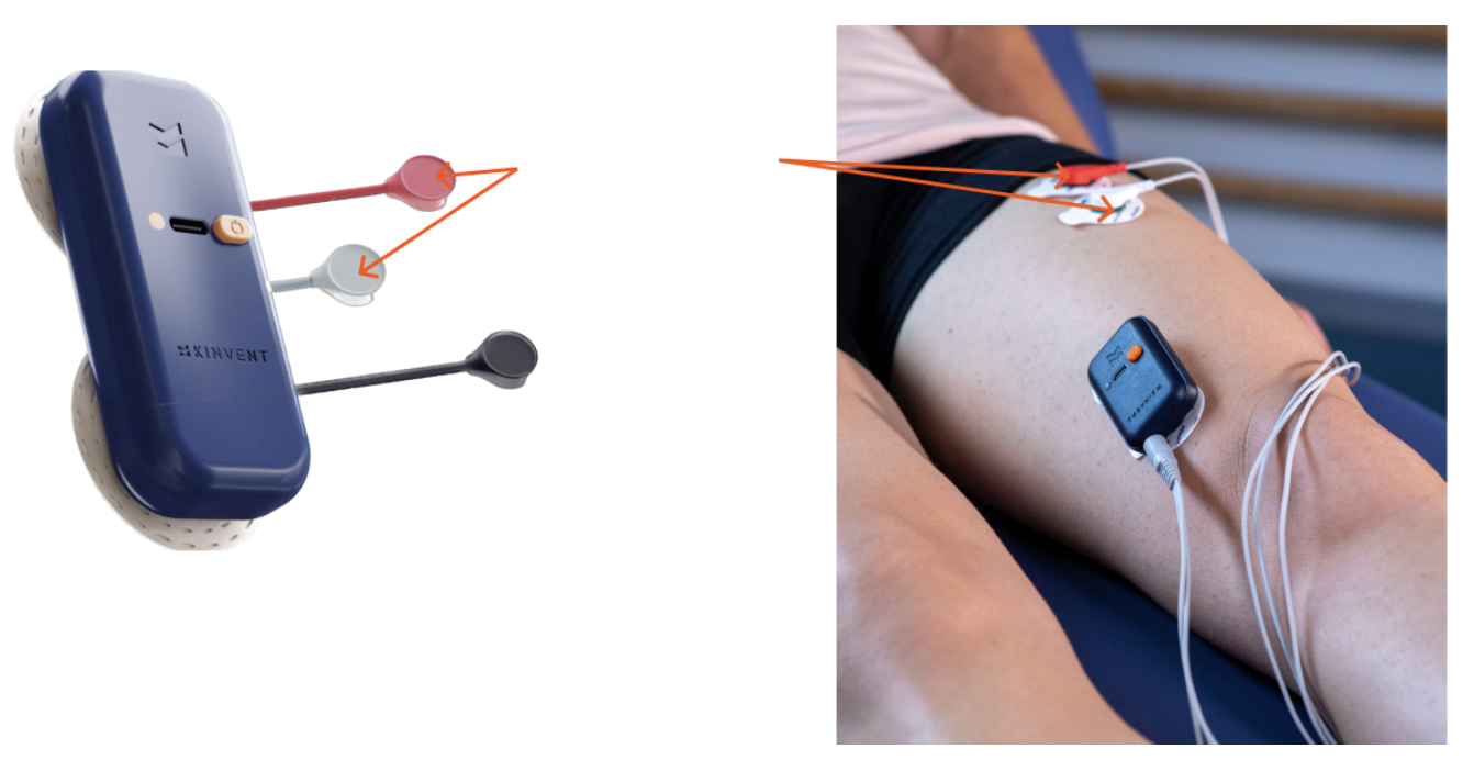
Correct placement of the electrodes in an electromyography (EMG) scan is essential to obtain accurate measurements of the electrical activity of the muscles.
Here is a guide on how to place the three electrodes of an EMG:
Electrode Placement
- Identify the muscle you wish to record.
- Place the first electrode (black - ground) on a non-active area of muscle, usually close to the area you are recording. This serves as a reference for the electrical activity.
- Place the other two electrodes (red and white) on either side of the area of interest.
Tips to Improve Recording Quality
- Ensure the chosen site is close to the targeted muscle mass to minimize the tissue layer between electrodes and muscle fibers.
- Position electrodes parallel to the muscle fibers to improve sensitivity and specificity. Electrodes placed perpendicular may reduce selectivity.
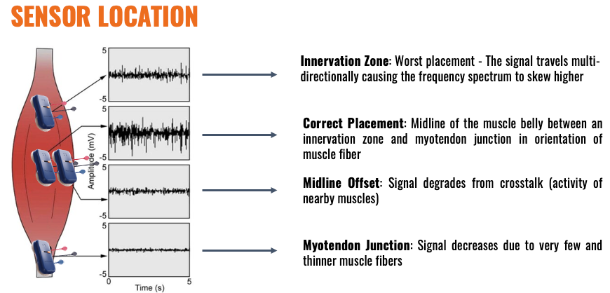
- Avoid the motor plate region. Electrodes placed here often record reduced amplitudes. The ideal position is in the middle of the muscle belly, between the innervation zone and the myotendinous junction.
- Select sites with clear anatomical landmarks to ensure consistency across sessions.
- Select sites that do not interfere with vision or movement, and avoid problem areas such as skin folds or bony prominences.
- Reduce interference from neighboring muscles by using appropriate electrode size and spacing.